Testicular Microlithiasis: What Your Ultrasound Shows
You went in for a scrotal ultrasound because of pain, swelling, or maybe a fertility check, and the radiology report came back with an unfamiliar phrase: testicular microlithiasis. For many men, this is the first time they have ever heard the term, and the immediate worry is the same — is this cancer?
Testicular microlithiasis explained in plain terms: it is the presence of tiny calcium deposits, called microliths, inside the seminiferous tubules of the testicle, visible on ultrasound as small bright spots. It is usually an incidental finding, it usually causes no symptoms on its own, and on its own — without other risk factors — it is generally not considered dangerous. This testicular microlithiasis explained article walks you through exactly what your ultrasound shows, how radiologists classify it, what the research says about cancer and fertility links, and what to do next.
Testicular Microlithiasis Explained: What It Actually Is
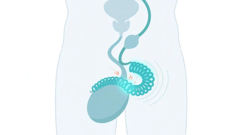
Testicular microlithiasis (pronounced tes-TIK-yoo-lar my-kroh-lih-THIE-uh-sis) refers to microscopic clusters of calcium salts that form inside the testicles. The deposits are composed of hydroxyapatite — the same mineral found in bone — surrounded by organic material. Each microlith measures roughly 1 to 3 millimeters across and sits inside the lumen of the seminiferous tubules, the tiny coiled tubes where sperm production takes place. To have testicular microlithiasis explained at the tubule level, researchers believe the deposits form when the Sertoli cells that line the tubules cannot fully clear away cellular debris, leaving a calcified core that builds up in concentric layers.
The condition itself causes no pain, no swelling, and no noticeable change you could feel. In the overwhelming majority of cases it is discovered by accident — a radiologist performing a scrotal ultrasound for some other reason, such as testicular pain, swelling, a fertility evaluation, or follow-up of a prior injury, notices the characteristic pattern.
Estimates suggest classic microlithiasis is found in roughly 0.1–0.5% of men in the general population, though it shows up more often — around 2–4% — in men who already have a clinical reason to undergo scrotal ultrasound. It is often bilateral, meaning both testicles are affected, though it can also be confined to one side.
What Your Ultrasound Report Is Really Showing
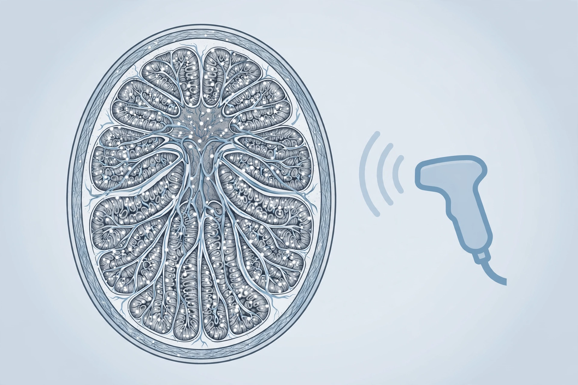
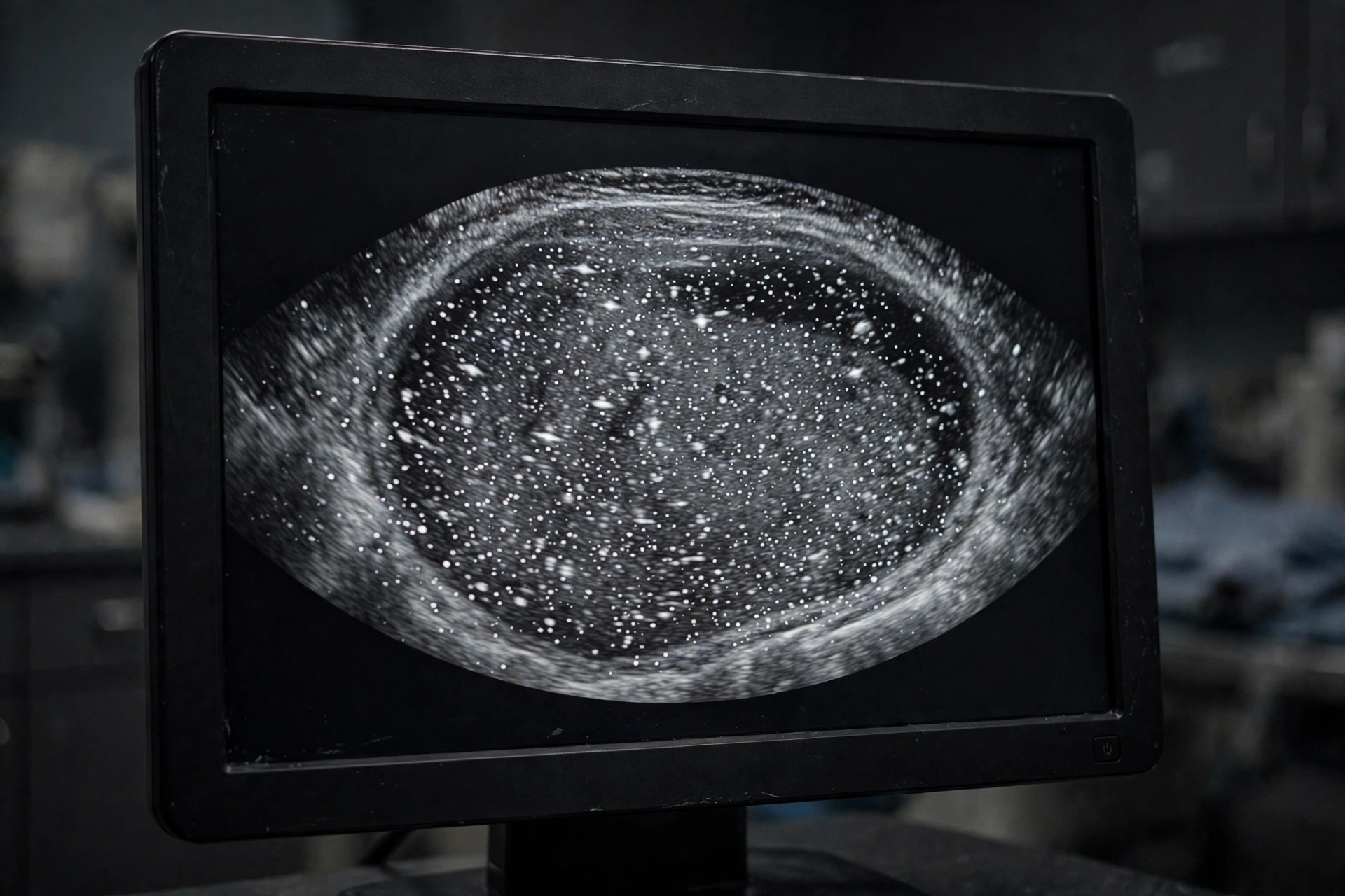
High-resolution scrotal ultrasound — typically using a probe of 7.5 MHz or higher — is the diagnostic method of choice, and it is the only imaging test needed. On the scan, microliths appear as discrete, bright (echogenic), punctate dots scattered through the testicular tissue, each 1–3 mm in size. Notably, they do not cast an acoustic shadow the way larger calcifications or stones do — that non-shadowing quality is one of the hallmarks the radiologist looks for.
Radiologists describe the pattern in a few recognizable ways:
- Scattered bright foci: small white dots distributed evenly through the testicular parenchyma, sometimes described as a “starry sky” or “snowstorm” appearance when dense.
- Symmetrical vs. asymmetrical: the dots are usually spread symmetrically through both testes; in about one in five cases the distribution is uneven, and some men have involvement of only one testis.
- Occasional artifacts: on color Doppler, the deposits may show a “twinkling” artifact, and comet-tail artifacts are sometimes visible — both are characteristic of tiny calcifications.
It is important to understand that an ultrasound diagnosis is descriptive, not diagnostic of anything else — and having testicular microlithiasis explained by your clinician in the context of your own scan is more useful than studying any single image. The scan shows the calcifications themselves; it does not show cancer. If the radiologist sees adjacent areas of abnormal tissue — for example, a focal darker (hypoechoic) area next to the dots — that is what would raise concern about a possible tumor, not the microliths alone. This is why the full report wording matters, and why your clinician reads it in the context of your history and physical examination.
How Radiologists Grade Testicular Microlithiasis
Not every finding is equal, and radiologists use a classification system to communicate how extensive the deposits are. The system most commonly referenced comes from the European Society of Urogenital Radiology (ESUR), which groups findings by the number of microliths visible per field of view:
| Grade / Category | What It Means | Typical Significance |
|---|---|---|
| Limited TML | Fewer than 5 microliths per field of view | Very common incidental finding; generally lowest concern |
| Classic TML | 5 or more microliths per field of view | Standard diagnostic threshold; risk depends on other factors |
| Diffuse TML (“snowstorm”) | Dense, widespread deposits across the parenchyma | Radiologist will look especially carefully for other abnormalities |
An older grading scheme (Backus et al.) divides findings into Grade 1 (5–10 per section), Grade 2 (10–20), and Grade 3 (more than 20), but the ESUR limited/classic/diffuse framework is what most modern reports follow. Why does this matter to you? When testicular microlithiasis is explained alongside its grade, the density of the finding helps your clinician decide on follow-up. A few scattered dots (limited) in an otherwise healthy man is generally managed very differently from dense bilateral deposits in a man with a history of undescended testicle or a prior testicular tumor.
The Cancer Question: What the Research Actually Says
This is the question every man asks first when testicular microlithiasis is explained to him, and it deserves a careful answer. Several studies do show a statistical association between testicular microlithiasis and testicular cancer — but the picture is more nuanced than the headline version. Here is the consensus as clinicians explain it, including guidance from Mayo Clinic specialists:
- The association exists mainly in selected populations. Most studies drew on ultrasounds ordered for another reason — pain, swelling, undescended testicle, infertility. In those groups, microlithiasis and testicular tumors appear together more often than chance would predict. A meta-analysis found an 8- to 10-fold higher chance of a concurrent germ cell tumor diagnosis in men who had microlithiasis plus other risk factors.
- In otherwise healthy men, the risk looks much lower. Studies of asymptomatic men show that microlithiasis is considerably more common than testicular cancer. Researchers therefore believe that microlithiasis by itself does not meaningfully raise cancer risk in a man with no symptoms and no other risk factors.
- Certain combinations warrant closer attention. Men with microlithiasis and infertility have been reported to carry a 10- to 40-fold higher tumor risk in some analyses (Barbonetti et al., 2019). Other red-flag combinations include bilateral deposits with an atrophic testis, a history of undescended testicle (cryptorchidism), or a prior germ cell tumor on the opposite side.
The practical takeaway: microlithiasis is not a diagnosis of cancer, and it is not a pre-cancer. It is a finding that prompts your clinician to review your overall risk profile. If that profile is clean, the standard advice is simply to practice regular self-examination and report anything new — the same vigilance recommended to every man. If additional risk factors are present, your clinician may recommend periodic ultrasound surveillance, typically every 1–2 years.
Testicular Microlithiasis and Fertility
The second most common concern is whether the calcium deposits interfere with fertility. The honest answer is that microlithiasis is frequently found in men being evaluated for infertility, but the direction of the relationship is not fully established. Some researchers view the deposits as a marker of disturbed tubule function — the same process that impairs sperm production may also leave calcium deposits behind — rather than as a direct cause of infertility. In other words, the microliths may be a signpost pointing at an underlying issue, not the issue itself.
What does this mean practically, once testicular microlithiasis is explained in a fertility context? If microlithiasis was found during a fertility workup and semen analysis results are abnormal, the finding supports a full reproductive evaluation rather than dismissal. Couples facing infertility can read more about common causes and when to seek specialist care in our guide to infertility causes and when to see a specialist. If your fertility testing is normal, the microlithiasis alone is not considered a reason to worry about future fatherhood, though discussing it openly with your clinician is always reasonable.
What Happens at Your Ultrasound Appointment
Many men arrive at the ultrasound already anxious, and not knowing what to expect makes it worse. A scrotal ultrasound is straightforward, painless, and takes about 15–20 minutes. Warm gel is applied, and a handheld transducer is moved gently over the area. There is no radiation, no needles, and no preparation such as fasting. Some men feel discomfort if the reason for the scan is a painful condition — that comes from the underlying issue, not the scan itself.
After the scan, a radiologist interprets the images and sends a written report to your clinician. Because findings like microlithiasis are incidental, it is common for men to learn about them days later at a follow-up appointment rather than on the day of the scan. Bring questions to that appointment: ask whether the finding was limited or classic, whether anything else in the report needs attention, and what surveillance, if any, your clinician recommends for your specific situation.

Self-Examination: Your Most Important Tool
Once testicular microlithiasis is explained to you, the practical next question is what you can do yourself. Across every guideline, the single most emphasized action for men with microlithiasis is regular testicular self-examination. Self-exams cannot detect the microliths themselves — the deposits are far too small to feel — but they can detect what matters most: a new lump, a change in size, a firm area, or persistent heaviness that could signal a developing tumor. Men who know their own baseline anatomy are the ones most likely to catch changes early.
The technique is simple: once a month, ideally during or after a warm shower when the skin is relaxed, roll each testicle gently between the thumb and fingers of both hands, feeling for anything new or different. One testicle is often slightly larger than the other, and that is normal — you are looking for change, not perfection. For a step-by-step walkthrough, see our testicular self-exam guide. If self-examination feels awkward at first, that is normal too; it becomes second nature within a few months.
It is equally important to know when pain or sudden swelling is an emergency rather than a routine concern. Testicular torsion — a twisting of the spermatic cord — causes abrupt, severe pain and needs treatment within hours. If you experience sudden intense pain, swelling, nausea, or a testicle sitting higher than usual, seek emergency care immediately, and review our guide to testicular pain causes and red flags so you know the warning signs in advance.
Treatment and Follow-Up: What Clinicians Typically Recommend
Once testicular microlithiasis is explained and identified, the next question is usually about treatment. There is no specific treatment for the condition itself — no medication dissolves the deposits, and surgery is not used for the condition. Management is entirely about monitoring and context:
- Healthy men with no risk factors: no routine surveillance ultrasounds are typically recommended; regular self-exams and routine medical care are the standard.
- Men with risk factors (undescended testicle history, prior tumor, testicular atrophy, significant infertility): clinicians often offer periodic ultrasound follow-up, commonly every 1–2 years, to watch for change.
- If something changes: a new lump, pain, or an abnormal area on imaging leads to the same workup any suspicious testicular finding would receive — physical examination, blood tests for tumor markers, and targeted imaging.
- Biopsy: reserved only for men at significantly elevated risk, per European urology guidelines — it is not a routine step for microlithiasis alone.
Because there is no drug or procedure to “fix” the finding, follow-up visits focus on reassurance, education, and keeping your personal risk profile up to date. If you change clinicians, make sure the microlithiasis finding is in your records so each new doctor has the full picture.
Frequently Asked Questions
Is testicular microlithiasis a type of cancer?
No. Testicular microlithiasis explained simply: it is a benign finding of microscopic calcium deposits in the testicular tissue. It is not cancer, not a pre-cancer, and not a tumor. The reason clinicians take it seriously is that some research links it to a higher chance of testicular tumors in men who also have other risk factors, so it prompts a careful review of your overall risk — but the finding itself is not a diagnosis of anything malignant.
Does testicular microlithiasis go away on its own?
No — the deposits are generally considered permanent and stable. They typically do not grow, spread, or resolve over time. Because they are stable and symptomless, they do not require treatment, and their presence alone does not cause damage to the testicle.
Can testicular microlithiasis cause infertility?
The condition is found more often in men being evaluated for infertility, but current evidence does not prove it directly causes infertility. It may instead be a marker of disturbed tubule function that accompanies other fertility problems. If your semen analysis is normal, microlithiasis alone is not considered a fertility concern; if you are having difficulty conceiving, a full reproductive evaluation is the appropriate next step regardless.
Should I get ultrasounds every year now?
Not necessarily. Routine serial ultrasound surveillance is generally recommended only for men who have microlithiasis plus additional risk factors — such as a history of undescended testicle, prior testicular tumor, testicular atrophy, or significant infertility. For men with no risk factors, guidelines do not call for scheduled repeat scans; self-examination and routine care are the usual plan. Your own clinician’s recommendation takes precedence, since they know your full history.
Can the microliths turn into kidney-stone-like stones?
No. Although they are made of calcium minerals like hydroxyapatite, testicular microliths are embedded in the tubule tissue and do not behave like urinary stones. They do not travel, do not cause blockages, and do not pass anywhere. Extremely rare case reports describe associated prostate calcification with stone passage, but this is not the natural course of ordinary testicular microlithiasis.
Does this affect my sex life or testosterone?
Testicular microlithiasis does not typically affect libido, erectile function, or testosterone production. It is a microscopic, localized finding that does not interfere with the hormonal functions of the testicle in the way that larger disease processes might. If you have symptoms such as low sex drive or erectile difficulties, they are almost certainly due to something else and deserve their own evaluation.
Is testicular microlithiasis hereditary?
There is no strong evidence that microlithiasis itself is directly inherited. It is associated with conditions that have genetic components, such as Klinefelter syndrome, but the calcium deposits are considered an acquired finding related to tubule function rather than a hereditary trait. Family history of testicular cancer remains worth mentioning to your clinician for independent reasons.
When should I call my doctor after this diagnosis?
Contact your clinician promptly if you notice a new lump or firm area, a change in the size or shape of either testicle, persistent pain or swelling, a feeling of heaviness, or fluid accumulation in the scrotum. Sudden severe testicular pain is an emergency — seek urgent care immediately, as that pattern suggests torsion rather than anything related to microlithiasis.
Medical Disclaimer
This testicular microlithiasis explained article is for educational purposes only and is not a substitute for professional medical advice, diagnosis, or treatment. Testicular microlithiasis affects each person differently depending on their individual risk factors, so the information here cannot replace a clinician who knows your history. Always consult a qualified healthcare professional about any concerns related to your testicular health, ultrasound findings, or fertility. Never disregard professional advice or delay seeking care because of something you have read here. If you experience sudden severe testicular pain or swelling, seek emergency medical attention immediately.